
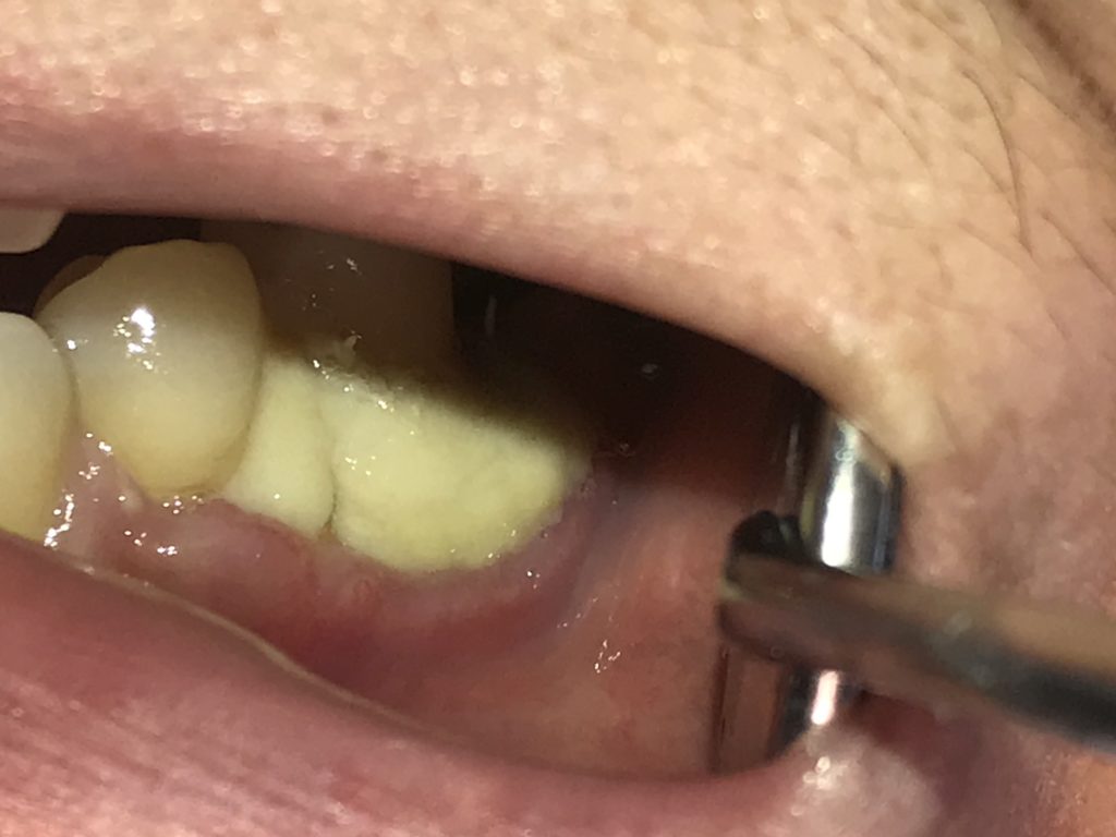
I did the soft tissue grafting on a patient with 3mm gingival recession on #35. I obtained a nice result when I finished the procedure that day with the gingiva covering the whole recession area.
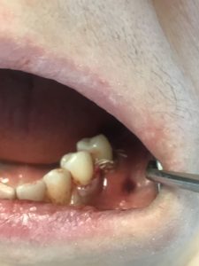
However, the patient came back 1 month after, complaining the gum-like graft was only left in the grafted area. I checked the surgical area and the alloderm was still stable, but the buccal gingiva for some reason was “opened up”. There was mild pain upon palpation but not pus coming out form the area.
I advised the patient to use warm salt water rinsing on that area, and re-evaluate in 1 month. I presricbed her clindiamycin 3 times per day for 1 week.
What have I done wrong? Is the alloderm supposed to be exposed? what treatment should i provide to the patient? will the gingiva “crawl up” the alloderm with time? Or do i need to remove the alloderm?
Of course it always starts with what the patient’s chief concern is, and what are his motivations for treatments and what are his expectations. In this case since a root coverage procedure will have significant limitations due to local anatomical factor that I will explain below, and there are no aesthetic demands, then simply restoring this area with composite would be my first course of action.
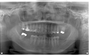
Where regards to the tooth that you have shown that you attempted root coverage for which is the last tooth in the arch number 35: Distal to it is an edentulous area where tooth number 36 was. With tooth number 36 being missing there has been significant resorption and remodeling of the Alveolus in that area and this contributed to the recession on the adjacent tooth number 35 that you’re trying to treat. The tooth may be slightly supererupted as well and Some of the “recession” may be a normal part of the enamel that has eroded.
The loss of alveolar interdental bone on the distal of 35 means that there is no support for coronally repositioning the flap with sling suture. The tissue will simply slide back on the bone towards its original position leaving the alloderm exposed.
Some of the alloderm will get integrated resulting in a thicker tissue biotype but with minimal or no root coverage. Also some of the exposed alloderm will simply disintegrate and fall away. Sometimes there is healing by secondary intention in these cases as the gingival marginal tissue crawls back to the tooth which serves to expand the zone of attached gingiva.
Also to be noted that there is an existing non-carious cervical lesion On tooth number 35 : Gingiva flaps margins don’t like to rest inside a concavity at the time of suturing. That’s not a stable position and a blood clot cannot form beneath the flap to stabilize the flap because the concavity prevents proper adherence of the flap to the tooth: so IN THE future to restore the enamel portion of the cervical defect first UP TO the CEJ before attempting root coverage and smooth Any rough root edges.
In this case I recommend to let the exposed alloderm heal naturally and reassess in another month. If there is no sign of infection such as redness heat swelling pain then just keep the Patient on chlorhexidine for another month.